Objectives: People living with HIV (PLWH) with low CD4 T-cell counts may be at a higher risk for severe coronavirus disease 2019 (COVID-19) outcomes and in need of efficient vaccination. The World Health Organization (WHO) now recommends prioritizing PLHIV for COVID-19 vaccination. Data on immune responses after messenger RNA (mRNA) vaccination in PLHIV in relation to CD4 counts are scarce. We aimed at assessing the humoral immune response in PLHIV after mRNA vaccination against COVID-19.
Methods: We examined a cohort of PLHIV after prime (n = 88) and boost (n = 52) vaccination with BNT162b2. We assessed levels of anti-severe acute respiratory syndrome coronavirus 2 (SARS-CoV-2) spike (S) protein-specific immunoglobulin G (IgG)/IgA and circulating neutralizing antibodies in plasma and correlated results to the cellular immune status. BNT162b2-vaccinated health care workers served as controls.
Results: All PLWH had a viral load of ≤ 200 HIV-1 RNA copies/mL and 96.5% had a viral load of < 50 copies/mL. Anti-S IgG and neutralizing antibody responses after BNT162b2 priming were significantly lower in PLHIV having a CD4:CD8 T-cell ratio of < 0.5. However, we observed robust humoral immunity in the majority of PLWH receiving antiretroviral therapy (ART) irrespective of CD4 T-cell nadir, current CD4 count or CD4:CD8 ratio after full BNT162b2 vaccination. Nevertheless, HIV-negative controls produced significantly higher mean anti-S IgG concentrations with less variability.
Conclusions: The majority of PLWH mounted robust responses after complete BNT162b2 vaccination but overall amounts of antibodies directed against the SARS-CoV-2 receptor-binding domain were variable. The impact on clinical efficacy remains unclear.
The majority of people living with HIV (PLWH) receiving effective antiretroviral therapy (ART) mount a functional adaptive immune response to severe acute respiratory syndrome coronavirus 2 (SARS-CoV-2) infection after predominantly mild coronavirus disease 2019 (COVID-19) disease.[1] HIV infection, however, has been shown to be an independent risk factor for both severe illness at hospital admission and in-hospital mortality.[2] SARS-CoV-2-specific T-cell responses are affected by the size of the naïve CD4 T-cell pool and the CD4:CD8 ratio in PLWH, in whom disparate antibody and T-cell responses have been observed.[1] In COVID-19, absolute numbers of lymphocyte subsets are differentially decreased according to clinical severity, with T-cell activation occurring in moderate but not severe disease.[3] The formation of T-cell memory is associated with disease recovery.[3] Some observational studies reported increased mortality in PLWH compared to HIV-negative individuals,[4–6] especially when CD4 T-cell counts were low,[7] but other studies did not find such an association.[8–10] Safe and efficient vaccination against COVID-19 is therefore of particular importance for PLWH, and the World Health Organization (WHO) now recommends prioritizing PLWH for COVID-19 vaccination.
For the ChAdox1 nCoV-19 (AZD1222) vaccine, efficient immune responses have been demonstrated in PLWH in independent clinical trials.[11,12] The phase 2 trial for BNT162b2 included 196 patients; however, data on CD4 counts were not included in the study.[13] A recent report on 14 PLWH demonstrated high antibody titres after two doses with varied titres after a single dose.[14] Another study demonstrated failure of seroconversion after two doses of BNT162b2 vaccine in a patient with uncontrolled HIV infection and a low CD4 count.[15]
We investigated whether a prime/boost vaccination regimen using BNT162b2 SARS-CoV-2 vaccine yields detectable anti-SARS-CoV-2 spike (S) antibody titres in PLWH on ART. We aimed to examine anti-S immunoglobulin G (IgG) responses in PLWH compared to those in seronegative controls and to assess whether CD4 T-cell nadir, current CD4 T-cell count or CD4:CD8 ratio was associated with the anti-S IgG response in PLWH.
Anti-S IgG was measured using an enzyme-linked immunosorbent assay (ELISA) (QuantiVac; Euroimmun, Lübeck, Germany) according to the manufacturer's instructions (dilution 1:500) and expressed as anti-S IgG concentration in RU/mL.[16] Anti-S IgA and anti-nucleocapsid (NCP) IgG measurements were performed according to the manufacturer's instructions (Euroimmun) and expressed as the IgA ratio (optical density divided by the calibrator). The cPass Neutralisation Antibody Detection kit (GenScript, Leiden, Netherlands) is a virus surrogate neutralization test (vSNT) and was used to detect circulating antibodies against SARS-CoV-2 (Wuhan) that block the interaction between the receptor-binding domain (RBD) of the viral spike glycoprotein and the angiotensin-converting enzyme 2 cell surface receptor. The sVNT shows good correlation to assays measuring viral entry inhibition.[17] For data analysis, GRAPHPAD PRISM 7 for Mac OS X was used (GraphPad Software, San Diego, CA, USA), employing linear regression analysis or the nonlinear exponential growth equation. Groups were compared by the Mann–Whitney rank sum test at a significance level of 0.05.
We assessed anti-S IgG, IgA and neutralizing activity in a surrogate virus neutralization test (sVNT) in the first 140 PLWH after vaccination with BNT162b2 at vaccination centres or by general practitioners. Between 8 March 2021 and 1 July 2021, we enrolled PLWH successively from our HIV outpatient clinic; 88 PLWH were tested after one and a further 52 PLWH after two doses of BNT162b2. Serology testing was performed on remaining plasma collected for routine CD4 T-cell measurement. All PLWH had a current viral load of ≤ 200 HIV-1 RNA copies/mL and 96.5% had a viral load of ≤ 50 copies/mL. All patients and controls had given written informed consent and the study was approved by the Internal Review Board of Hannover Medical School (approval no. 2411–2014 and 8973_BO_K_2020). Four PLWH with evidence for previous SARS-CoV-2 infection as determined by anti-SARS-CoV-2 NCP IgG were excluded from the analysis. BNT162b2 prime and boost were administered a mean of 29 days (range 21–47 days) apart. As the time-points of vaccination were not predefined and serology testing was performed during the next routine HIV care visits after vaccination, the mean time of anti-S IgG, anti-NCP IgG and inhibition measurement was 18.7 days (range 0–42 days) after the first and 35 days (range 1–128 days) after the boost vaccination. Patient characteristics are listed in Table 1.
Healthy controls with unknown HIV status, no major comorbidities and no previous COVID-19 were from our COVID-19 Contact (CoCo) Study, in which we assess immune responses in health care workers after BNT162b2 vaccination as part of the vaccination campaign at our university hospital.[13]
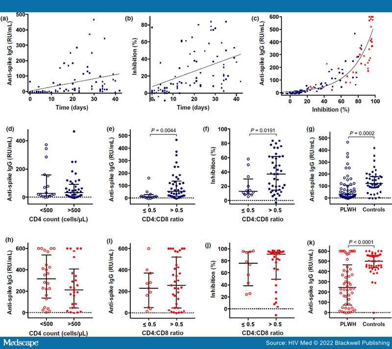
Anti-S IgG became detectable in most patients from day 10 after BNT162b2 priming (Figure 1a), and increasing anti-S IgG concentrations were accompanied by increasing inhibitory activity in the vSNT, suggesting generation of neutralizing antibodies (Figure 1b). Both parameters reflecting functional humoral immune response were closely correlated within the entire cohort after prime and boost vaccination (n = 129; r[2] = 0.8479; Figure 1c). To assess the impact of the cellular immune status in PLWH on early anti-S humoral responses after vaccination, we restricted our analysis to plasma taken at least 10 days after priming. Comparison of PLWH (n = 56) with CD4 counts below and above 500 cells/μL revealed no significant differences for anti-S IgG [median anti-S IgG 26.23 RU/mL; n = 16; interquartile range (IQR) 148.1 RU/mL vs. 33.35 RU/mL; n = 40; IQR 86.78 RU/mL, respectively; Figure 1d] or IgA (data not shown). Similarly, the CD4 nadir was not associated with anti-S IgG or inhibitory activity. However, individuals with CD4:CD8 ratio > 0.5, compared with those with CD4:CD8 ratio ≤ 0.5, had significantly higher anti-S IgG (median 53.3 RU/mL; n = 42; IQR 120.2 RU/mL vs. 11.47 RU/mL; n = 12; IQR 26.3 RU/mL, respectively; P = 0.0044; Figure 1e) and IgA (data not shown). Similarly, titres of neutralizing antibodies assessed in the vSNT were three times as high in PLWH having a CD4:CD8 ratio > 0.5 compared with the other group (median 37.03%; n = 42; IQR 46.8% vs. 12.66%; n = 12; IQR 21.3%, respectively; P = 0.0191; Figure 1f). A similar difference was observed when patients were categorized in groups with CD4 cell counts above and below 25% (data not shown), suggesting that CD4 T-cell immunity is associated with humoral vaccine-induced immunity in the early phase after priming.
Figure 1.
Humoral immune responses after prime (blue) and boost (red) with BNT162b2 in people living with HIV (PLWH). (a) Anti-severe acute respiratory syndrome coronavirus 2 (SARS-CoV-2) spike (S) immunogobulin G (IgG) (RU/mL) over time after BNT162b2 priming. (b) Percentage inhibition assessed by virus surrogate neutralization test (sVNT) over time after BNT162b2 priming. (c) Correlation of anti-S IgG with sVNT after priming. (d–g) > 10 days after prime vaccination: anti-S IgG vs. current CD4 T-cell count (d) and CD4:CD8 ratio (e) categories; (f) percentage inhibition vs. CD4:CD8 ratio categories; (g) anti-S IgG of PLWH vs. HIV-negative controls. (h–k) After secondary boost vaccination: anti-S IgG vs. current CD4 count (h) and current CD4:CD8 ratio (i); (j) neutralizing antibodies vs. CD4:CD8 ratio; (k) comparison of PLWH with controls. The lines represent median and interquartile range.
Boost BNT162b2 vaccination diminished associations with the CD4:CD8 ratio, as anti-S IgG (Figure 1h–j), inhibitory activity and IgA (not shown) responses in PLWH were comparable irrespective of their current CD4 count, CD4 T-cell percentage (data not shown) or CD4:CD8 ratio. Confirming the overall robust humoral immune response, most PLWH had high titres of neutralizing anti-S antibodies a mean of 35 days (range 1–128 days) after complete BNT162b2 vaccination. However, five PLWH (12%) had low neutralizing activity (< 20%) indicating inferior humoral responses (Figure 1i). When comparing anti-S IgG responses to the response after BNT162b2 prime/boost in health care workers (all anti-NCP IgG negative) at a mean of 26 days (range 18–37 days) after complete vaccination, responses in PLWH were more variable and significantly lower after prime (median 31.51 RU/mL; n = 56; IQR 101 RU/mL vs. 119.8 RU/mL; n = 41; IQR 92.37 RU/mL in health care workers; P = 0.0002; Figure 1g) and especially after boost (median 246.2 RU/mL; n = 50; IQR 218.7 RU/mL vs. 502.5 RU/mL; n = 41; IQR 118.8 RU/mL in health care workers; P < 0.0001; Figure 1k).
In summary, our preliminary data demonstrate robust humoral immune responses after complete BNT162b2 vaccination in PLWH on ART irrespective of their CD4 T-cell nadir and current CD4 T-cell immune status, suggesting that mRNA vaccines are immunologically suitable for this patient group. Importantly, the high variability and significantly lower humoral anti-S IgG response in comparison to HIV-negative controls may indicate inferior immunity in PLWH. However, the magnitude of reduced vaccine-induced immune responses in PLWH may be partially explained by differences in the sex and age distribution or comorbidities between the two groups, as we were unable to fully match for these parameters. Data presented here and our previous observations in healthy vaccinees support the conclusion that PLWH should be encouraged to get their vaccine schedules completed in order to reach a high anti-S IgG titre and inhibitory activity.[17] Associations of overall cellular immunity in PLWH and S-specific IgG were only evident early after priming and seemed not to be predictive for the overall response. Larger studies will be required to identify factors with predictive value for inferior vaccine responses in PLWH, such as lack of neutralizing antibody responses. The contribution of vaccine-elicited cellular immune-mediated protection against COVID-19 is still unclear and we propose more comprehensive functional CD4 and CD8 T-cell analyses in PLWH to assess the relationship between ART-induced immune reconstitution and protective anti-S immunity.
Our study was preliminary and has several limitations. We were unable to collect data on reactogenicity and studied only one mRNA vaccine. The available seronegative control group did not perfectly match the PLWH cohort in demographic characteristics, leading to possible bias. We did not quantify neutralization against variants of concern such as the alpha, beta, gamma or delta variant, which may be more relevant in the future. Our sample size was too small to assess the impact of lower CD4 counts (< 200 cells/μL), ART regimens, age, sex or comorbidities on vaccine responses. In addition, we assessed only surrogates for protection with yet unknown relevance for infection and disease prevention, and longer follow-up will be required to gain information on long-term immunity and protective efficacy over time. Finally, it will be interesting to determine adaptive T-cell immunity in PLWH related to their immune reconstitution during ART, as cellular immunity may significantly contribute to the vaccine-mediated protection against COVID-19. For now, continuation of COVID-19 vaccination with BNT162b2 is warranted and robust humoral immunity can be expected in the majority of PLWH on ART. However, because of the preliminary evidence suggesting lower humoral immunity in PLWH compared to HIV-negative controls, PLWH may become a target population for prioritization to receive a second booster vaccination.
Funding information
European Regional Development Fund; German Center for Infection Research
Acknowledgements
This project received funding from the European Regional Development Fund (Defeat Corona, ZW7-8515131). GMNB is supported by the German Center for Infection Research (TTU-HIV 04.820 and 08.821). We thank Margot Zielinska-Skowronek, Birgit Heinisch, Janine Topal, Kerstin Sträche, Melanie Ignacio, Annika Heinemann, Janne Vehreschild, and Melanie Stecher for technical and logistical support. Open access funding enabled and organized by ProjektDEAL.
Ethical Approval
The study was approved by the Internal Review Board of Hannover Medical School (approval nos 2411–2014 and 8973_BO_K_2020).
HIV Medicine. 2022;23(5):558-563. © 2022 Blackwell Publishing