Case Description
An 11-year-old male child presented with snapping of right wrist since 6 years of age. Initially asymptomatic, it became worrisome due to associated pain with movement.
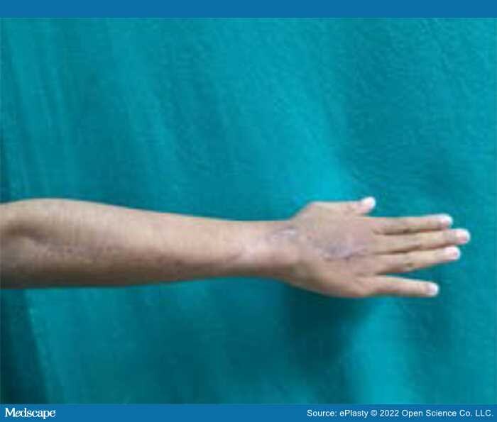
Clinically, a subcutaneous cord-like structure was palpable on wrist extension, extending from the dorsoulnar aspect of distal right forearm across the wrist to the metacarpophalangeal joint of middle, ring, and index fingers (Figure 1). Snapping was elicited with palpable click on wrist flexion due to volar migration of anomalous cord across the ulnar styloid process (Video1).
Figure 1.
Examination showing band over the extensor aspect of ulnar side of right wrist
Magnetic resonance imaging (MRI) revealed well-defined linear structure posteromedial to extensor carpi ulnaris tendon appearing isointense to tendons, likely representing accessory tendon.
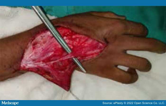
On exploration, a dense cord was found subcutaneously at the wrist above the extensor retinaculum extending to the metacarpophalangeal joint of the middle finger with accessory slips to the ring and index fingers (Figure 2). Proximally this cord-like structure originated from deep fascia just distal to lateral epicondyle with no muscular origin (Figure 3). All extensor tendons were found in respective compartments under the extensor retinaculum with normal function. The abnormal cord was excised and sent for histopathological study.