Bacterial and Viral Coinfection in Idiopathic Pulmonary Fibrosis Patients

The Prevalence and Possible Role in Disease Progression

Mohsen Moghoofei; Shayan Mostafaei; Nasim Kondori; Michelle E. Armstrong; Farhad Babaei

Disclosures

BMC Pulm Med. 2022;22(60) 

In This Article

Results

Study Subjects

A final diagnosis of IPF was made after multidisciplinary team discussion; 12 of the 79 recruited patients did not fulfill the American Thoracic Society (ATS) diagnostic criteria for IPF and were subsequently excluded from the study.[2] The remaining 67 subjects with IPF were predominantly men 38 (56.7%) with the mean age 62.8 (SD = 12.44) years. Twelve (17.9%) and 55 (82.1%) of patients were stable and acute exacerbation (AE-IPF). The median time from diagnosis to an acute disease status (AE-IPF) was 90 days. All IPF patients suffered chronic pneumonia, as diagnosed by CT scan in recent years. Nine of 55 (16.3%) of the AE-IPF patients had both fever and myalgia, which was suggested viral-like illness. All patients had moderately severe disease at enrollment as characterized by carbon monoxide diffusing capacity (DLCO) (70.5% predicted ± 3.96), Forced Expiratory Volume in 1 s (FEV1) (70.9% predicted ± 4.30) and Forced Vital Capacity (FVC) (75.3% predicted ± 4.37). Additional details are included in Table 1.

Prevalence of Bacterial-, Viral- and Co-infection Rates in IPF Patients

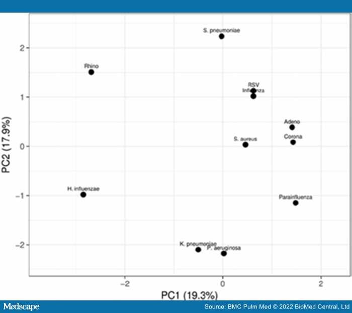
Of the 67 samples collected, 12 (17.9%) samples were positive for viral infection, 7 (10.4%) samples were positive for bacterial infection and 40 (59.7%) samples were positive both viral and bacterial infection (coinfection). Among the mono infections, rhinovirus (39.7%) and H. influenza (46%) were detected at the highest rate in IPF patient samples. In contrast, coronavirus (14.3%), S. aureus (25.4%) and K. pneumoniae (25.4%) were detected at the lowest rate in IPF patient samples. Additional details are included in Table 1.

Effect of Bacterial-, Viral- and Co-infection on Pulmonary Function in IPF Patients

Here, we carried out an analysis of the effect of viral-, bacterial- and co-infection on pulmonary function in IPF patients. In Table 2, we examined FVC, FEV1 and DLCO indices in IPF patients who were non-infected (Group 1), infected with bacteria only (Group 2), infected with virus only (Group 3) and coinfected with virus and bacteria (Group 4). In this study, there was a significant decrease in FVC values (% predicted) in IPF patients who were coinfected with virus and bacteria compared with patients were non-infected (p = 0.013; Table 2). There was also a significant difference in DLCO values (% predicted) in IPF patients who were infected with bacteria (p = 0.030; Table 2) or coinfected (p = 0.001; Table 2), respectively, compared with non-infected IPF patients. There was no significant difference in FEV1 values (% predicted) in non-infected (Group 1) compared with any of the infected groups of IPF patients (Groups 2, 3 and 4).

Effect of Bacterial-, Viral- and Co-infection in on Disease Status, Survival Status and Survival Time on IPF Patients at 60-month Follow-up

Three important factors including disease status (AE-IPF versus stable-IPF), survival status (death vs. survive) and survival time (months-to-death) were investigated in non-infected, viral infected, bacterial infected and coinfected IPF patients (Table 3). In this study, we demonstrated that IPF patients who were coinfected had more unstable disease, had a higher incidence of death and a short survival time compared with non-infected IPF patients. Specifically, a significantly greater percentage of coinfected IPF patients (55%) were found to be in the AE phase of disease compared with non-infected patients (0%) (p < 0.001; Table 3). This suggests that virus and bacterial coinfection led to an increase in the severity of the disease. Investigation of the number of deaths within the IPF study cohort revealed that a significantly greater percentage of coinfected IPF patients (37.5%) died compared with non-infected patients (0%) (p = 0.043; Table 3). An investigation of the survival time from diagnosis (months-to-death) in IPF patients demonstrated that survival time in coinfected patients (32.9 ± 9.12 months) was significantly less than survival in non-infected IPF patients (42.5 ± 6.55 months) (p = 0.026; Table 3). Furthermore, Figure 1, using Kaplan–Meier survival curve analysis and the log-rank test, we demonstrated that IPF patients who were co-infected (blue line) had a significantly increased risk of mortality compared (Log rank text: p = 0.031) with IPF patients who were non-infected (black line) [Hazard ratio: 8.12; 95% confidence interval (CI) 1.3–26.9].

Figure 1.

Kaplan–Meier survival curve analysis for comparison of time until death (in months) between uninfected (black line) and bacterial—(red line), viral—(green line) and co-infected (blue line) IPF patients. Coinfected IPF patients had a significantly increased risk of death compared with uninfected patients [Log rank test: p = 0.031; Hazard ratio: 8.12; 95% CI 1.3–26.9]

Principal Component Analysis (PCA) of Uninfected, Viral-, Bacterial- and Co-infected IPF Patients

Based on Kaplan–Meier curves and log-rank test, we established that IPF patients who are coinfected with virus and bacteria have significantly reduced FVC and DLCO (% predicted), an increased rate of AE-IPF, an increased incidence of death and risk of mortality, and a reduced survival time from months-to-death from diagnosis, respectively, compared with non-infected IPF patients. Here, we employed principal component analysis (PCA), based on the first and second components of the IPF patients included in this study in order to investigate the pattern of bacterial and virus infections, and coinfection in these patients. (Figure 2). In the PCA score plot, the viral infections (1) RSV and influenza and (2) adenovirus and coronavirus have the most similar coinfection patterns. In the context of bacterial infection, K. pneumonia and P. aeruginosa have the most similar co-infection pattern.

Figure 2.

Principal component analysis (PCA) score plot depicting the relationship between bacterial-, viral- and co-infection groups according to PCA based on the first and second principal components for n = 67 IPF patients

Analysis of Longitudinal Decline in FVC in Uninfected, Viral-, Bacterial- and Co-infected IPF Patients Over a 60-month Follow-up Period

Analysis of FVC change (% predicted) from baseline was carried out over a 60-month period post-diagnosis as a predictor of IPF disease progression and mortality risk (Figure 3). We observed a significant decline in FVC change in IPF patients with bacterial infection (p < 0.001), viral infection (p < 0.001) and co-infection (p < 0.0001) compared with uninfected patients. Furthermore, the decline in FVC was significantly greater in co-infected IPF patients compared with patients infected with bacteria (p < 0.001) or virus only (p < 0.001).

Figure 3.

Time-trend analysis of FVC changes from baseline to 60 months after IPF diagnosis. p value (uninfected vs. coinfection) < 0.0001. p value (uninfected vs. viral infection) < 0.001. p value (uninfected vs. bacterial infection) < 0.001. p value (viral infection vs. bacterial infection) = 0.679. p value (coinfection vs. viral infection) < 0.001. p value (coinfection vs. bacterial infection) < 0.001

processing....