Hand Tumors: An Individual Surgeon's Retrospective Review

Kamil Amer, MD; Katie Otero MD; Brendan Smith MD; Ramazi Datiashvili MD

Disclosures

ePlasty. 2022;22(e1) 

In This Article

Abstract and Introduction

Abstract

Background: Tumors of the hand are encountered frequently and represent a variety of pathologic diagnoses, both benign and malignant. Even within a single pathologic type, presentation can vary. This study reviews hand tumors encountered by an individual surgeon and described presenting features to better aid in clinical decision making.

Methods: A retrospective chart review of patients presenting with a hand tumor between January 2005 and December 2017 from an individual surgeon's perspective was performed. Pertinent data were extracted by researchers and statistical analysis was completed with GraphPad Prism (GraphPad Software, Inc).

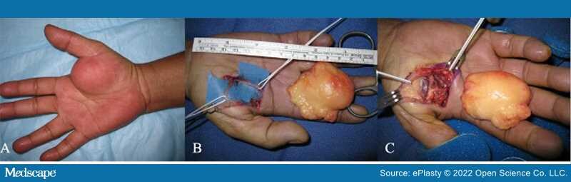
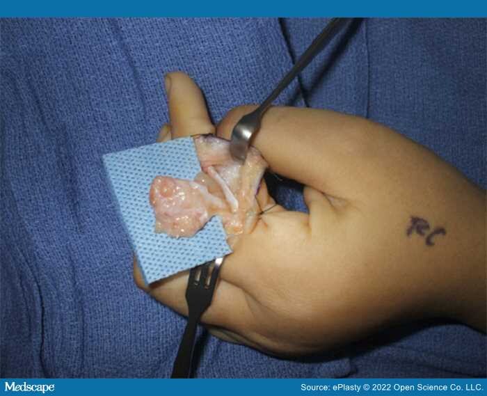
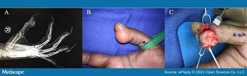
Results: A total of 101 patients aged 14 months to 87 years (mean age, 40.52 years) were included. Within this patient group, soft tissue tumors accounted for 97%, malignant neoplasm 2%, and bone tumors 1%. Ganglion cysts were most common (54.5%) followed by hemangiomas (9.9%), giant cell tumors (6.9%), granulomas (5.9%), and fibromas (5%). A total of 54.5% of patients reported pain and 43.5% reported decreased range of motion (ROM).

Conclusions: In this patient cohort, ganglion cyst was the most common tumor type and presented with pain and deficits in ROM. This is contrary to the asymptomatic presentation of such cases in the literature. Other common tumors were hemangiomas, giant cell tumors, granulomas, and fibromas. Malignant tumors were rare, as expected, and both cases presented without pain or tenderness.

Introduction

Tumors of the hand are encountered frequently by physicians and can be difficult to accurately identify due to the wide variety of differential diagnoses. Although an estimated 95% of hand tumors not involving the skin are benign, malignant tumors are possible and must be identified for proper treatment.[1] Distinguishing between benign and malignant lesions on clinical presentation alone, however, can be difficult.

Ganglion cysts are the most common benign tumors of the hand, with a higher predominance in women and often occurring in the second through fourth decades of life.[2] These lesions are frequently located in the dorsal wrist (70%) and less frequently in the volar wrist (20%), though appearance in the tendon sheaths and distal interphalangeal (DIP) joints has also been reported.[3] Median and ulnar nerve compressions have been reported which can cause pain and functional deficits.[4]

There are several other benign tumors that may occur in the hand. Lipomas are well-defined, soft, subcutaneous nodules that may present anywhere within the hand. (Figure 1) While generally asymptomatic, lipomas present in the carpal tunnel, Guyon's canal, or deep palmar space may result in nerve compression.[5] Neurofibromas which are benign tumors of the nerve sheath, may raise suspicion for genetic conditions such as neurofibromatosis or, if multiple are present, Von Recklinghausen's disease. (Figure 2) These conditions increase the risk of malignant transformation.[6,7] Osteochondromas are most commonly found in the proximal phalanx and show the presence of bony exostosis on radiographs.[9] (Figure 3) Although malignant degradation to chondrosarcoma may occur elsewhere in the body, this has not been described with lesions of the hand.[4,8]

Figure 1.

Lipoma of the right thenar eminence in 57-year-old female on inspection (A) and during dissection and resection (B, C)

Figure 2.

Plexiform neurofibroma resection in the left index finger of a 12-year-old female

Figure 3.

Osteochondroma of the right thumb in a 48-year-old female on radiograph (A), inspection (B), and during resection (C)

Malignant tumors, although less common, are also possible in the hand. They can arise from a variety of different areas including cutaneous tissue, bone, and connective tissue. The most common is chondrosarcoma, a malignant soft tissue tumor derived from cartilage-producing cells which may arise as a primary or secondary lesion.[9] This tumor can be seen in adults over 60 years of age, most commonly in the proximal phalanges, specifically of the fifth digit.[8] They present as a chronic, painful swelling that is poorly defined on radiograph.[8,10] Metastasis is very rare (<10%) with the most common site being the lungs. A computerized tomography scan of the chest may be warranted if metastatic spread is suspected.[8]

This retrospective review is a single surgeon's perspective on these complex and varying hand tumors. The purpose is to better understand how these tumors present in a specific population of patients. Variables assessed include patient demographics, tumor type, and presenting symptoms. Understanding the features of hand tumors in this cohort of patients may further guide treatment for these complex cases in the future.

processing....