Riluzole, a Glutamate Modulator, Slows Cerebral Glucose Metabolism Decline in Patients With Alzheimer's Disease

Dawn C. Matthews; Xiangling Mao; Kathleen Dowd; Diamanto Tsakanikas; Caroline S. Jiang; Caroline Meuser; Randolph D. Andrews; Ana S. Lukic; Jihyun Lee; Nicholas Hampilos; Neeva Shafiian; Mary Sano; P. David Mozley; Howard Fillit; Bruce S. McEwen; Dikoma C. Shungu; Ana C. Pereira

Disclosures

Brain. 2022;144(12):3742-3755. 

In This Article

Results

Sample Characteristics and Demographics

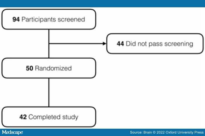
A total of 94 participants were screened at the two performance sites, of which 44 did not meet inclusion/exclusion criteria. The remaining 50 participants were randomly assigned to receive riluzole (n = 26) or placebo (n = 24). Of these, 22 patients receiving riluzole and 20 patients receiving placebo completed the study and had both FDG-PET time points. The diagram in Figure 2 shows the participant disposition.

Figure 2.

Enrolment, randomization and trial completion.

Enrolled participants comprised 26 females and 16 males, aged 58 to 88, and 58% were apolipoprotein 4 (APOE ɛ4) carriers (Table 1, one participant unavailable). There were no significant between-group differences in baseline characteristics of the patients with respect to age, sex, education or APOE ɛ4, although there was a trend for a greater proportion of APOE ɛ4 carriers in the riluzole group. Baseline neuropsychological measures were well balanced for MMSE, NPI, ADL total, Clinical Dementia Rating scale total in riluzole group in comparison to placebo; however, the riluzole group trended as more impaired in ADAS-cog than placebo at baseline (P = 0.08; Table 1).

Neuroimaging Outcome Measures
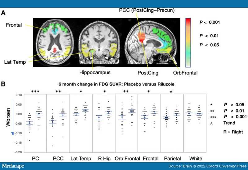
FDG-PET. The study's main primary outcome measure confirmed a difference between arms in FDG-PET cerebral metabolic changes over the 6-month treatment period, with less decline in multiple prespecified brain regions in the riluzole group in comparison to placebo group.

There were no significant or trend-level differences between study arms at baseline in the regional SUVRs that were compared, or in the FDG Alzheimer's disease progression score. Given the trend-level difference between study arms in APOE ɛ4 dose, analyses were performed and compared with and without its inclusion as a covariate. Supplementary Table 3 presents the mean, standard deviation and significance findings for the FDG-PET comparisons. PC glucose metabolism, a primary end point, was significantly preserved in the riluzole-treated group in comparison to placebo over the 6-month period [effect size (d) 1.31; P < 0.0002] with APOE ɛ4 dose included as covariate, P < 0.0003 without APOE ɛ4 dose included, with the effect significant using any of several different reference regions (paracentral P < 0.0002, centrum ovale P < 0.008, whole brain P < 0.016, cerebellar cortex P < 0.03) (Figure 3A–C). PC significance readily survived Bonferroni correction for multiple comparisons. Regional cerebral glucose metabolism was more preserved in the riluzole group in comparison to placebo in several other prespecified regions of interest including precuneus (P < 0.007, d = 0.84), lateral temporal (P < 0.014, d = 0.80), right hippocampus (P < 0.025, d = 0.72) and frontal cortex (P < 0.035, d = 0.67), and the exploratory subregions of orbitofrontal cortex (P < 0.008, d = 0.86) and PC-precuneus subregion (P < 0.007, d = 0.88) (Figure 4). Most of these still showed trend-level significance if corrected for multiple comparisons. Age, sex, education and APOE ɛ4 dose were not significant contributors to treatment effect. No differences were observed in control regions such as subcortical white matter, pons and cerebellar vermis.

Figure 3.

Comparison of changes in FDG-PET in PC over 6 months. (A) PC region of interest (representative sagittal slice) in FDG-PET. (B) Comparison between placebo and riluzole-treated arms of the absolute and percentage change in PC FDG SUVR over the 6-month treatment period. (C) Individual change from baseline to follow up in PC SUVR in placebo (left) and riluzole (right) treated arms. (D) Comparison of change in PC SUVR by APOE ɛ4 carrier and non-carrier subgroups, and by younger and older age groups. Individual values are shown with mean and standard error bars.

Figure 4.

Comparison of changes in FDG-PET in prespecified regions of interest over 6 months. (A) Region of interest boundaries shown in representative slices, colour-coded to indicate the significance levels in comparisons between placebo and riluzole-treated arms of the 6 month change in FDG SUVR. (B) Comparison between placebo and riluzole-treated arms of the 6 month change in FDG SUVR for posterior cingulate (PC = PostCing), combined PC and precuneus (PCC), lateral temporal (LatTemp), right hippocampus (Hip), orbitofrontal (OrbFrontal), frontal, parietal and subcortical white matter (as a comparator, expected to remain stable). For each region, data-points are shown for the placebo group on the left and for the riluzole-treated group on the right. Individual values are shown with mean and standard error bars.

When groups were stratified and analysed separately on a post hoc basis (using non-parametric tests due to subgroup size) by APOE ɛ4 carrier status, age and sex, the treatment effect of riluzole group having less decline than placebo was observed in both APOE ɛ4 carriers and non-carriers [P < 0.004 in carriers (n = 8 placebo, 15 riluzole, effect size 1.526) and P < 0.09 in non-carriers (n = 11 placebo, 7 riluzole, d = 0.89), in both younger and older groups (P < 0.002 in older group, n = 13 placebo, 15 riluzole, d = 1.370 and P < 0.08 in younger group, n = 7 placebo and seven riluzole, d = 0.96); Figure 3D], and in males and females (both groups P < 0.02; n = 14 placebo, 12 riluzole in female group and n = 6 placebo, 10 riluzole in the male group). Inclusion of APOE ɛ4 dose in the by-age group analysis increased the P-value for study arm to 0.13 in the younger group, with APOE ɛ4 dose showing a trend-level influence in this age group (P < 0.07) but not the older age group (P < 0.78). Inclusion of APOE ɛ4 dose in the by-sex group analysis decreased the P-value for study arm to P < 0.008 in the female group, with APOE ɛ4 dose showing a trend-level influence in the female group (P < 0.09) but not the male group (P < 0.84).

FDG-PET measures have been shown to correlate with cognitive decline and predict disease progression.[62–64] The exploratory FDG-PET Progression Classifier score analyses showed a trend of less increase (less worsening) in Alzheimer's disease progression score in the riluzole group in comparison to placebo (P < 0.07; Figure 5A). There was a trend-level greater difference between arms in APOE ɛ4 carriers than non-carriers. FDG-PET Alzheimer's disease progression scores correlated with ADAS-cog at baseline (all participants R = 0.61, P < 0.00002; placebo group R = 0.57, P < 0.008; riluzole group R = 0.48, P < 0.0004) and changes in FDG Alzheimer's disease progression scores correlated with changes in ADAS-cog (all participants R = 0.46, P < 0.002; placebo group R = 0.56, P < 0.011; riluzole group R = 0.29, not significant and reduced range of score increases) (Figure 5B) over the 6 months of the study. Additional correlations were observed between FDG-PET and cognitive measures as shown in Figure 6, including relationships between baseline FDG Alzheimer's disease progression score and MMSE (R = 0.61, P < 0.00002; Figure 6A), FDG PC SUVR and MMSE (R = 0.35, P < 0.00003; Figure 6B), lateral temporal SUVR and ADAS-cog (R = 0.54, P < 0.0002; Figure 6C) and orbitofrontal SUVR and NPI score (R = 0.52, P < 0.0004; Figure 6D). The robust correlations observed between FDG-PET brain metabolism and cognitive measures in our dataset support our secondary outcome measure related to neuropsychological assessment.

Figure 5.

Comparison of FDG-PET progression score changes over 6 months. (A) Comparison between placebo and riluzole-treated arms of the change in FDG progression score. (B) Correlation between Alzheimer's disease progression score at baseline and ADAS-cog score at baseline (left) and between the 6-month change in Alzheimer's disease progression score and in ADAS-cog (right) for all study participants.

Figure 6.

Correlations between FDG-PET measures and cognitive performance. Correlations at baseline between: (A) FDG Alzheimer's disease progression score and MMSE score; (B) posterior cingulate-precuneus (PCC) score and MMSE score; (C) lateral temporal FDG SUVR and ADAS-cog score; and (D) orbitofrontal FDG SUVR and NPI score.

1H MRS

Voxel Tissue Composition and Spectral Quality. The proportions of grey matter, white matter or CSF in the PC or the hippocampal voxels did not differ between groups. Except for an inconsequential increase in the full width at half maximum of the PC water resonance between baseline (4.5 Hz) and 6 months (6 Hz), the spectral quality parameters were remarkably stable, with no group differences in the unsuppressed reference tissue water signal (W) or signal-to-noise ratios observed as a function of time in any of the voxels. The voxel tissue composition and spectral quality control data are summarized in Supplementary Table 2.

Posterior Cingulate 1H MRS Results

As a primary end point, neither NAA/W nor NAA/tCr showed a temporal change (group × visit interaction: P = 0.14 or P = 0.89, respectively) (Supplementary Figure 1). 1H MRS results in secondary end point PC glutamate (Glu) levels showed a group × visit interaction when expressed as Glu/tCr (P = 0.05) and a trend-level interaction when expressed as Glu/W (P = 0.3), with both levels increasing after the first 3 months of treatment (Figure 7A). No significant changes from baseline to 6 months were observed. In post hoc analyses, within the riluzole group NAA/W and Glu/W levels were correlated positively for all three time points, while NAA/tCr and Glu/tCr were positively correlated at baseline (Figure 7B). Within the placebo group, both NAA/W versus Glu/W and NAA/tCr versus Glu/tCr were positively correlated for all three time points (Figure 7B). 1H MRS measures of Glu/W correlated positively with MMSE and negatively with ADAS-cog across all participants (Figure 7C). Across all participants Glu/tCr positively correlated with MMSE (P = 0.01) but not ADAS-cog (P = 0.39).

Figure 7.

Comparison of changes in 1 H MRS measures. (A) 1H MRS measures of Glu/W (top) and Glu/tCr (bottom) levels changes in PC at baseline, 3 and 6 months. (B) Correlations at baseline, midpoint and end point between NAA/W and Glu/W in riluzole and placebo groups (top) and NAA/tCr and Glu/tCr in riluzole and placebo groups (bottom). (C) Correlations at baseline between Glu/W and MMSE (top) and Glu/W and ADAS-cog (bottom) across participants.

Hippocampal 1H MRS Results

On an exploratory basis, a significant increase in GABA/W was observed in the left hippocampus with a significant group × visit interaction (P = 0.03) (Supplementary Figure 2B) and as a trend level in GABA/tCr (P = 0.3). Left hippocampus GABA/W levels positively correlated with memory performance; see Logical Memory 1 and 2 testing correlations at baseline in Supplementary Figure 2C. No significant changes were seen in any measure of Glx or NAA levels in right or left hippocampus nor GABA levels in right hippocampus.

Neuropsychological testing was a secondary measure for correlation with neuroimaging biomarkers, although the study was not powered for a significant neuropsychological effect. Trend-level findings are shown in Supplementary Figure 3 for the key functional measures of ADL Inventory, NPI and ADAS-cog.

Adverse Events

There were no statistical differences in adverse events between treatment groups, with 23 of 26 patients (88.5%) in the riluzole group and 22 of 24 (91.7%) in the placebo group having at least one adverse event during the study. Serious adverse events occurred in 2 (7.7%) in the riluzole group and 1 (4.2%) in the placebo group. The most common side effects in the riluzole group consisted of abdominal discomfort (15.4% in riluzole, none in placebo); diarrhoea (15.4% in riluzole, 8.3% in placebo); dizziness (15.4% in riluzole, 4.2% in placebo); urinary frequency (11.5% in riluzole and none in placebo), nausea (7.7% in riluzole, none in placebo), cough (19.23% in riluzole, 12.5% in placebo), elevated liver enzymes (7.7% in riluzole and 4.2% in placebo) and others (Supplementary Table 1). Among the randomized patients, 4 of 26 (15.4%) in the riluzole group and 3 of 24 (12.5%) in the placebo group had an adverse event that led to removal from the trial. There were no significant differences in the frequency of participants who were discontinued from the trial due to adverse events.

processing....