Managing Diffuse Peripheral Ischemia in Antiphospholipid Syndrome: Anticoagulation, Immunosuppression, or Vasodilation?

Caroline H. Siegel, MD; Doruk Erkan, MD, MPH

Disclosures

April 14, 2021

This article originally appeared on Hospital for Special Surgery's website.

Case Report

A 39-year-old man with a history of well-controlled type 1 diabetes (T1D) since childhood and migraine presented with 1 week of pain and cyanosis of the fingers of the left hand and toes of both feet. Although he had no history of Raynaud phenomenon, warm temperature yielded transient improvement. He had primary antiphospholipid syndrome (APS), diagnosed 6 years prior on the basis of unprovoked lower-extremity deep vein thrombosis and persistent antiphospholipid antibodies (positive lupus anticoagulant test, high-titer anticardiolipin antibody [aCL] immunoglobulin [Ig] G, and high-titer anti-beta2-glycoprotein I antibody [aβ2GPI] IgG).

He was on warfarin (target international normalized ratio [INR]: 2.5-3), aspirin 81 mg daily, atorvastatin 10 mg daily, and verapamil 240 mg daily. Two weeks prior to presentation, his INR had increased to 7 while on antibiotics for sinusitis; it nadired at 1.3 a week later after warfarin was held.
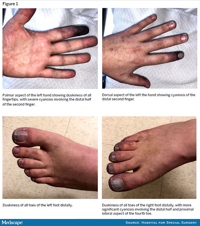
Figure 1.

On physical examination, he had distal cyanosis in the left hand and both feet (Figure 1). Laboratory test results were notable for platelet count of 94 K/μL (normal: 150-450 K/μL), INR of 1.8, one-time troponin elevation to 0.19 ng/mL (normal: ≥ 0.04 ng/mL), and elevated serum transaminases, which peaked at 3 times the upper limit of normal and then trended down over 1 week. Serum levels of complement components 3 and 4 were normal, aCL IgG was > 150 GPL, and aβ2GPI IgG was > 150 SGU. Antineutrophil cytoplasmic antibodies were negative. Glycosylated hemoglobin (A1c) was 6.8% (normal: ≤ 5.6%). Transthoracic and transesophageal echocardiography did not identify valvular vegetations. Magnetic resonance angiography (MRA) of the left hand revealed severe occlusive arterial disease with extensive collateralization (Figure 2).

Figure 2.

Due to concern for APS-associated microvascular disease evolving to catastrophic APS (CAPS), he was started on methylprednisolone 125 mg twice daily, hydroxychloroquine 200 mg twice daily, and intravenous (IV) heparin. Aspirin and atorvastatin were continued; verapamil was switched to nifedipine 60 mg daily. Four days after admission, rituximab 1 g IV was given (repeat dose 2 weeks later). We initiated sildenafil 20 mg three times daily, with partial improvement in the cyanosis of the left second finger.

One week later, the patient underwent a left upper-extremity nerve block with marked but temporary benefit. Intravenous immunoglobulin (IVIG) 2 g/kg was administered over 5 days and platelet count normalized. The patient also received IV epoprostenol with no immediate response; this was discontinued after 12 hours due to flare of migraine, and sildenafil was increased to 40 mg three times daily.

By the time of discharge, methylprednisolone had been tapered to 10 mg twice daily and warfarin was resumed. Aspirin, atorvastatin, hydroxychloroquine, nifedipine, and sildenafil were continued. The pain and cyanosis in the left hand had resolved except in the gangrenous distal second finger; his toes remained dusky.

Discussion

Patients with APS may experience a spectrum of thrombotic manifestations: moderate to large vessel disease, microvascular disease, and CAPS. Our patient presented with ischemic digits due to microvascular disease and unexplained mild transaminase and troponin elevations but did not meet CAPS classification criteria.

While anticoagulation is the mainstay of treatment for moderate to large vessel thrombosis in APS, microvascular APS and CAPS require additional modalities. First-line therapy for CAPS includes anticoagulation, glucocorticoids, IVIG, and/or plasma exchange. For microvascular APS and CAPS, adjunctive therapies include statins and hydroxychloroquine. Rituximab may be effective in patients with microvascular manifestations. The optimal management of peripheral ischemia in APS is not well established.

Our patient's clinical presentation was atypical. Microvascular APS leading to diffuse peripheral ischemia is rare, especially without meeting CAPS classification criteria. Longstanding T1D probably contributed to the chronic arterial disease seen on MRA. Although he had no known T1D-related microvascular complications, peripheral microvascular disease can develop before retinopathy and may not correlate with the degree of glycemic control. Prior infection and subtherapeutic INR level may have triggered this patient's acute presentation. Vasospasm seemed to play a role as well, perhaps causing steal phenomenon in the context of underlying arterial occlusions. Thus, in addition to anticoagulation and immunomodulation, we optimized his calcium channel blocker, added a phosphodiesterase-5 inhibitor, and pursued trials of a prostaglandin and chemical sympathectomy.

In conclusion, for this complex case of refractory peripheral ischemia in a patient with APS and T1D, we used an aggressive multimodal treatment strategy to maximize perfusion to multiple digits at risk for infarction.

Comments

3090D553-9492-4563-8681-AD288FA52ACE
Comments on Medscape are moderated and should be professional in tone and on topic. You must declare any conflicts of interest related to your comments and responses. Please see our Commenting Guide for further information. We reserve the right to remove posts at our sole discretion.

processing....